How Does Caffeine Affect Your Brain Function?


brain and coffee

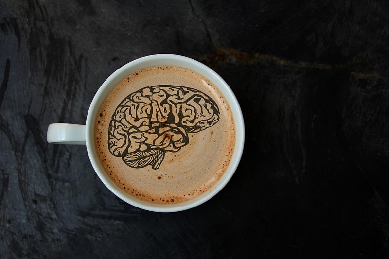
Every day, billions of people drink caffeine. Their reasons for this differ. Either they are relying on it to wake up, or to get through an afternoon slump, or maybe a night shift.

Caffeine is a well-known natural stimulant and is one of the most widely-used ingredients globally (1).

One of the main reasons caffeine is popular is its negative effects on the brain, including anxiety and sleep. But then, several studies have commended its many health benefits.

This article examines the effects of caffeine on your brain, as well as how it affects your sleep and your health in general.

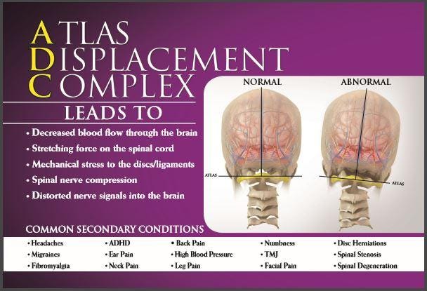
How caffeine can affect your nervous system.

What is Caffeine?

It is a natural stimulant widely found in coffee, cacao, and tea plants. It stimulates the central nervous system and the brain and helps you to remain alert while also preventing fatigue.

According to history, the very first tea was brewed in 2737 BC (1).

It was discovered by a shepherd in Ethiopia who observed that it gave his flock extra energy. And then, in the late 1800s, caffeinated soft drinks came on board, followed by energy drinks.

Currently, over 80% of the global population takes at least one caffeinated product each day. In North America, over 90% of adults take caffeinated products daily (1).

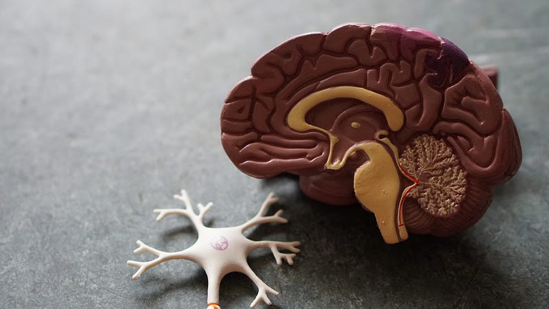
How Does Caffeine Affect Your Brain?

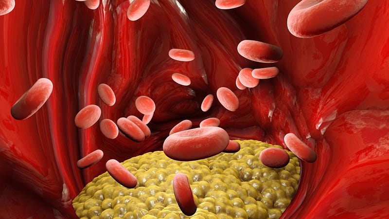
After caffeine is consumed, it is absorbed from the digestive system into the bloodstream. Once in the bloodstream, it moves to the liver and is metabolized (broken down) into elementary compounds that can affect your organs and how they function.

However, the main effect of caffeine occurs in the brain. Caffeine blocks adenosine’s effects. Adenosine is a neurotransmitter that causes your brain to relax and gives you that tired feeling (2).

Normally, the adenosine levels in your body build up over time, causing you to feel tired and give you that sleepy feeling.

But caffeine will keep you mentally alert and awake by binding to adenosine receptors but without activating them. This will block adenosine’s effects, thus reducing tiredness (3).

Caffeine also increases the level of adrenaline in the blood and the activities of norepinephrine and epinephrine in the brain (3).

This stimulates the brain and triggers a state of alertness, focus, and arousal. Caffeine is classified as a psychoactive drug because of its potential to affect your brain. Also, caffeine is “quick-acting.” For example, the amount of caffeine present in a cup of coffee can reach your bloodstream within 20 minutes, while its full effects can be felt within an hour of taking it (1).

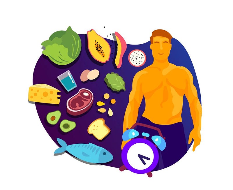
Foods that Contain Caffeine

Caffeine is found naturally in nuts, leaves, or the seeds of certain plants. These sources are then harvested and processed, resulting in the production of caffeinated beverages and foods.

Below, we have the amounts of caffeine you may get per 240ml serving of some beverages (1):

  • Coffee: 102–200mg
  • Espresso: 240–720mg
  • Energy drinks: 50–160mg
  • Yerba mate: 65–130mg
  • Chocolate milk: 2–7mg
  • Cocoa beverage: 2–7mg
  • Decaffeinated coffee: 3–12mg
  • Soft drinks: 20–40mg

You can also find caffeine in some foods. For example, an ounce (28g) of milk chocolate contains 1–15mg, while an ounce of dark chocolate will provide 5–35mg (4).

Caffeine is also present in some over-the-counter drugs or prescription drugs like pain, allergy, and cold medications. It is also used in weight loss supplements.

Ice, Ice, Maybe?


Ice — The Illusionary Treatment for Inflammation

The practice of applying ice to an injury to reduce inflammation has been used for decades. The thing is, there’s ZERO peer-reviewed scientific evidence to support this practice. Recent studies have shown that ice may affect the healing process.

The question is, “Why do many healthcare providers use ice on an injury to help reduce inflammation?”

The idea behind this practice is that ice impedes blood circulation to the injured area and also minimizes swelling. But is this right? We’ll learn more from this article.

There’s nothing bad about inflammation

Inflammation is not bad — that’s a fact. It is a normal aspect of human physiology, so is there any reason why it should be stopped? You see, whenever a tissue is injured, the body makes attempts to remove the damaging stimulus and trigger the healing process.

The healing process includes increasing the circulation of blood and the flow of lymph to and from the affected area. This enhances the movement of inflammatory cells and nutrients to the injured site. On the other hand, the lymphatic system flushes out the waste and fluid that has accumulated due to inflammation.

Let’s talk about inflammation

The human body uses two systems of circulation. The major one is the blood circulatory system which includes the heart, arteries, and veins.

The lymphatic system is the second one. It comprises small bags that channels fluid to the heart. One thing about the lymphatic system is that it doesn’t have a pumping mechanism.

What powers the movement of lymphatic fluid is the activity and movement of the surrounding muscles. Muscle activity causes a squeezing effect that pushes the fluid towards the direction of the heart. If there’s no movement, the swelling will remain and will not be evacuated from the injured area.

Does the application of ice to inflamed tissues work?

Well, ice slows down the flow of blood to the injured site. Of course, this will reduce the pain by numbing pain receptors but the swelling may not readily be removed.

Because there is no movement with ice (zero muscle activity), a backflow of swelling occurs in the lymphatic system. So, rather than the inflammation decreasing, it increases due to poor evacuation.

What’s the right way to treat injuries and inflammation?

Whenever you are injured, you should put on an active approach. Doing so will provide the best benefit. A proactive approach mustn’t be complicated. It could be as simple as some gentle movement, usually pain-free. This will maintain movement and function.

There’s a suitable acronym for the proactive approach, and it is METH.

METH stands for Movement, Elevation, Traction, and Heat and was recently coined by a Toronto-based trainer and exercise physiologist, John Paul Catanzaro.

Movement

Because the lymphatic system hasn’t got a self-pumping mechanism, it relies on muscle movement to push the fluid. The implication is that getting rid of swelling does require some movement. If you experience pain while moving a limb, then you can apply a muscle stimulator to the affected muscles. This will promote muscle activity, and enhance lymph flow without causing pain.

Elevation

On its own, it may be the least effective due to its passive nature. On the other hand, combining elevation with movement will enhance the flow of lymph away from the injured area. The results can be very dramatic.

Traction

Traction depressurizes the injured area. Combining traction with movement promotes healing because it boosts the flow of fresh blood to the affected area. Also, combining traction with movement will reduce pain as it depressurizes the injured area.

Heat

This is a simple one. Heat enhances circulation. So, technically, it will boost the flow of fresh blood to the affected area. Fresh blood supplies fresh oxygen which speeds up the healing process.

Conclusion — Ice is not nice for injuries

With everything taken into account, we can accept that a harmony between the RICE and METH strategies is significantly relying upon the seriousness of the injury, area harmed, and span/time-pass following the injury.

For example, ice can help with diminishing torment and possibly important if the usage of natural inflammatories or painkillers are not available.

Basically, we should be liberal in our way to deal with treating delicate tissue wounds. What works for one individual, may not function admirably for another person.

Next time you think RICE, consider the METH strategy for treating that delicate tissue injury.

Can You Build Muscle While Doing Intermittent Fasting?


Does intermittent fasting affect lean muscle mass?


Intermittent fasting
has been in the news lately. This is not surprising as it is a very popular diet. But does it affect lean muscle mass?

There are several types of intermittent fasting, but they all share one thing in common — which is that they last longer than the regular overnight fast.

Several pieces of nutrition-based research have shown that intermittent fasting can help a person to lose some fat. However, there’s a little bit of alarm in that it seems to have the potential to cause muscle loss.

In this article, we will discuss explicitly how intermittent fasting affects your lean muscle mass.

What is intermittent fasting?

As popular as the concept of intermittent fasting is, there’s still some confusion as to what it is.

This is not surprising because intermittent fasting is a really broad term that describes several kinds of eating. The commonest types of intermittent fasting include (1):

  1. Time-restricted fasting: Intake of calories is restricted to a couple of hours daily, ranging from 4–12 hours. In most cases, there’s always an 8-hour eating period.
  2. Alternate-day fasting: In this form of fasting, you will alternate between non-fasting and fasting days. What this implies is that you’ll fast on other days, but not fast on some. Some people may not have any meals on fast days, but in many cases, people do have small meals on fasting days.
  3. 5:2 diet: It is similar to periodic and alternate-day fasting. On a 5:2 diet, you’ll eat well for 5 days a week, and for the remaining 2 days, you’ll have just 25% of your normal calories.
  4. Periodic fasting: Another name for this is whole-day fasting. In a periodic fast, you will have occasional fasts separated by weeks or days of normal eating.

What happens during weight loss?

Can you fast and gain muscle at the same time?

You see, weight loss involves the shedding of unwanted body fat, but it is much more than that. When you lose weight, you equally lose lean body mass, visceral fat, bodily fluids, and of course, water weight.

Does intermittent fasting affect lean muscle mass?

Water weight refers to the fluids that can accumulate in your body tissues due to stress, poor nutrition, or hormonal changes. Instead of the fluid getting released, it is stored by your body between your skin and organs. This water weight may be referred to as bloating.

When you start intermittent fasting, your carb levels will reduce and your body will switch to glycogen for energy. Glycogen is activated by water. So, your body makes use of the water in its stores to create energy from glycogen.

Fat burning starts immediately after your water bloat is drained. This process also burns your muscle. Research suggests that you can burn fat and also gain lean muscle simultaneously during an intermittent fast.

What are the odds of losing muscle when fasting?

Most studies on intermittent fasting have been targeted primarily at weight loss (1).

Note that one can lose weight without exercise, but in this case, the weight loss will be due to the loss of lean mass and fat. Leans mass means everything (including muscle) except fat (2).

This applies to weight loss caused by intermittent fasting as well as other diets.

Many studies have shown that a person may lose small amounts of lean mass after fasting intermittently for several months (1).

But then, other studies could not find any loss of lean mass (3, 4).

Many studies, however, have concluded that intermittent fasting may help maintain lean mass as compared to other non-fasting diets. But there’s a need for further research on this subject (5).

Generally, you may not lose your muscle on an intermittent fast compared to other weight-loss diets.

Is an Aspirin a Day Good or Bad for Your Heart?

Photo Credit: https://stock.adobe.com/images/daily-aspirin/86009033 © Sherry Young


Have you ever suffered a stroke or a heart attack? No doubt you may have taken some low-dose aspirin as a remedy. But is daily aspirin use a healthy choice?

In this article, we will discuss the shocking side effects that a daily dose of aspirin has on your heart health.

Shaking up the Salt and High Blood Pressure Myth


salt sodium chloride
Photo by Charles Deluvio on Unsplash


We’ve received several warnings about the dangers of salt to our health.

Many health organizations have claimed that high salt intake causes several health conditions, like heart disease and high blood pressure. But then, these claims do not have strong scientific backing despite decades of research (1).

Also, many nutritional studies have shown that not eating enough salt can cause harm to your health.

This article will differentiate the myths and the facts about salt (sodium) and high blood pressure.


What is salt?

Another name for salt is sodium chloride. The salt that we use in cooking is made of 40% sodium and 60 % chloride.

Salt is a major dietary source of sodium. This explains why the words “sodium” and “salt” are used interchangeably.

There are also trace amounts of potassium, calcium, zinc, and iron in some varieties of salt. Table salt contains iodine as well (2, 3).

Salt contains essential minerals that serve as electrolytes. These electrolytes regulate fluid balance, muscle function, and nerve transmission.

Most foods contain salt, naturally. Salt is also added to foods as a flavoring agent.

It served as a preservative in the past. High amounts of salt can stop bacteria from growing in food.


Does sodium affect blood pressure?

Well, several health authorities have warned against excessive intake of sodium. We’ve been advised to take no more than 2,300mg of sodium daily, or preferably less (4, 5, 6).

That’s the equivalent of one teaspoon of salt (or 6 grams).

But the bad news is that over 90% of adults in the United States consume more than the recommended amount of salt (7).

Excess consumption of salt is believed to raise blood pressure, thus increasing the risk of stroke and heart disease. However, this may not be so true.

Yes, consuming less salt can lower blood pressure, mostly in people who have salt-sensitive hypertension (8).

The average reduction of salt is however very subtle for healthy individuals. According to a 2013 study, restricting salt intake in people with normal blood pressure caused only a 2.42 mmHg reduction in systolic blood pressure and a 1.00mmHg reduction in diastolic blood pressure (9).

That’s the same thing as going down from 130/75 mmHg to 128/74 mmHg. Is this worth the stress of eating a tasteless diet? I don’t think so.

Also, several review studies suggest that no evidence reducing your salt intake will reduce your risk of stroke, heart attacks, or death (10, 11).


A healthy diet is a key to lowering blood pressure

A healthy diet plays an important role in lowering blood pressure and keeping it at optimal levels. Studies have shown that adding greens and other healthy foods to your diet tones down your blood pressure (12, 13).

Fatty fish, berries, leafy greens, beans, seeds, lentils, carrots, and citrus fruits can help you achieve optimal blood pressure levels.

Leafy green vegetables contain lots of nitrates. Nitrates are known to help regulate blood pressure. Studies have shown that taking 1–2 servings of vegetables rich in nitrates daily can lower high blood pressure for at least 24 hours.

High blood pressure is about a potassium insufficiency — not necessarily a sodium excess.

Leafy greens that can help ease your blood pressure include:

  • Collard greens
  • Cabbage
  • Spinach
  • Mustard greens
  • Kale
  • Fennel
  • Swiss chard

green smoothie receipe
Photo credit: https://www.100daysofrealfood.com/green-smoothie-recipe/


There’s no better way to eat your daily greens than stirring your spinach into stews and curries, sauteing your Swiss chard with some garlic (makes a great side dish), or baking some kale chips.

You can also blend them together and make a tasty green smoothie out of it like the one pictured above. What’s your favorite type of green smoothie?

Ligament Laxity: A Missed Diagnosis from a Car Crash


woman experiencing neck pain from a car crash.
Photo Credit: Paolese

Ligament laxity is also known as ligamentous instability. It is a condition that causes chronic pain and may affect any joint in the body. It is important to note that ligament laxity consists of those joints that extend beyond their normal range of motion.

Now, according to the American Academy of Orthopaedic Surgery, the commonest soft tissue injuries occur in or around the ligaments, tendons, and muscles.

Sudden trauma causes acute injuries. Sudden trauma includes a twist, fall, or sharp blow to any part of the body.

Examples of acute injuries include:

  • Contusions
  • Sprains
  • Strains

What are some other types of ligament laxity?

It is also important to note that there are other soft tissues in the body that are prone to injury. This includes the heart, lungs, brain, or other organs in the body. But medically, though, soft tissue injuries are usually limited to the tendons, ligaments, and muscles.

A closer examination of the structure of ligaments, tendons, and muscles shows that they’re basically connective tissues. The National Institute of Health defines connective tissues as “the material inside your body that supports many of its parts.” It shapes your tissues and keeps them strong.


What causes ligament laxity?

Ligament laxity results from extreme flexibility of the ligaments that surround your joints. The extreme flexibility of the ligaments allows these joints to move beyond their normal range of motion. Ligament laxity in most individuals is genetic and may start at a young age. At least one joint may be affected, and in some cases, the entire body (known as general joint hypermobility).

Ligament laxity also occurs after a ligament is damaged and fails to heal properly. The affected ligament loosens or becomes lax and does not support the joint as well as it should. In severe cases, joint damage may cause ligament laxity especially when the joint moves beyond its normal position. If ligament instability occurs in the region of the spine, the patient may suffer disc degeneration or osteoarthritis. Shoulder dislocations and sprained ankles are also common occurrences associated with ligament instability.

According to a 2013 study by Hauser and colleagues, the structure of an injured ligament is replaced with tissue that is similar to scar tissues biochemically, biomechanically, and grossly. Scar tissues that have been fully remodeled remain microscopically, grossly, and functionally different from normal tissues. Because the remodeled ligament tissue is inferior to normal ligaments, there’s a resulting laxity of the ligament, leading to disability of the affected joint, while other soft tissues surrounding the joints are predisposed to further damage.


Ligament laxity caused by automobile accidents

You see, most car crashes cause soft tissue damage. This soft tissue damage in turn causes ligament laxity.

Using whiplash as a case study, the torso and the head move in directions opposite to the impact force — this causes damage to the ligaments in the neck and spine.

Photo by C Joyful on Unsplash

This may result in other injuries, as well as chronic pain. Secondary conditions experienced may include:

  • Numbness
  • Strains and sprains
  • Swollen joints
  • Pains in the neck and back
  • Discomfort when moving the torso or the neck

Diagnosis of ligament laxity is done via stress radiographs, joint mobility, and symptom evaluation. Treatment may vary but usually include Foundational Correction, anti-inflammatory medication, pain medication, braces, physical therapy, and/or movement, elevation traction, and heat (METH). In extreme cases, surgery of the neck or back may be recommended.


Can ligament laxity lead to permanent damage?

Ligament laxity can affect a person’s work-life or ability to carry out his/her activities of daily living.

Truth be told, ligament laxity is a costly condition, with expensive medical bills and even loss of one’s source of livelihood.

Also, ligamentous instability may lead to other injuries. The emotional distress resulting from the accident may trigger anxiety, depression, or mental health issues.

What’s more? Ligament laxity may have long-lasting physical effects if left uncorrected.


References

Ellis SJ. Tremor and other movement disorders after whiplash-type injuries

Journal of Neurology, Neurosurgery & Psychiatry 1997;63:110–112.

Derrick, L J, and B M Chesworth. “Post-motor vehicle accident alar ligament laxity.” The Journal of orthopaedic and sports physical therapy vol. 16,1 (1992): 6–11. doi:10.2519/jospt.1992.16.1.6

Cerebral Palsy and Foundational Correction


Photo Credit: By piter2121
Photo Credit: By piter2121

Cerebral palsy (CP) is more than one condition. The term cerebral palsy describes a group of conditions sharing similar symptoms. It is a neurological condition that usually shows up during infancy, leading to body movement and muscle coordination issues. The symptoms of cerebral palsy may be due to brain damages or abnormalities to the brain that affects the child’s ability to maintain the correct posture, balance, or movement.

Does cerebral palsy have specific symptoms? If yes, what are they? Can the nervous system function be improved? Is there a way to restore the body’s ability to control the muscles? We’ll learn about this and more in this article.


What is cerebral palsy?

Cerebral palsy (CP) is a group of conditions that affect the muscles' coordination and movement. In most cases, hearing, sensation, and vision are affected.

When you hear the word cerebral, what comes to your mind? Well, cerebral refers to the brain. “Palsy,” on the other hand, means “problems” with the movement of the body or “weakness.”

Most childhood motor disabilities are caused by cerebral palsy. The Centers for Disease Control and Prevention (CDC) report shows that globally, 1.5–4 of every 1000 children are affected by this condition.

Symptoms of cerebral palsy

The symptoms of this condition vary. It may be mild in some people and very severe in others. Some cerebral palsy patients may have difficulty sitting and walking. Other cerebral palsy patients may find it hard to grasp objects.

The severity of the symptoms reduces over time. Symptom severity also depends on which part of the brain was affected.

Photo by Robina Weermeijer on Unsplash

Common signs of CP include:

· Stiff muscles (spasticity)

· Involuntary movements of tremors

· Variations in muscle tone

· Late attainment of motor skill milestones, like sitting up alone, crawling or rolling over.

· Difficulty walking

· Difficulty swallowing or excessive drooling

· Neurological conditions, like intellectual disabilities, blindness, and seizures

· Lack of muscle coordination (ataxia)

It is important to note that many children are born with cerebral palsy. However, the symptoms only show up after some months or years. The symptoms usually show up before the child attains the age of 3–4.

You should consult your doctor if you think that your child has cerebral palsy. Effective treatment depends a lot on early diagnosis.

What causes cerebral palsy?

Well, cerebral palsy is mostly caused by injury to the brain or abnormal development of the brain. Damage to the developing brain can affect the brain region that controls posture, coordination, and body movement.

Damage to the brain usually occurs before the child is born. However, it may also happen during the first year of life. Most of the time, the exact cause isn’t known.

Photo Credit: irissca
Photo Credit: irissca

Factors that may cause cerebral palsy include:

· Jaundice in the child

· Genetic mutations that cause poor development of the brain

· Maternal infections, like herpes simplex or German measles

· Asphyxia neonatorum (lack of oxygen during delivery)

· Head injuries due to falls, automobile accidents, or child abuse

· Bleeding into the brain (intracranial hemorrhage)

· Brain infections, like meningitis and encephalitis.

Who is at risk for this condition?

Several factors may increase a child’s risk for cerebral palsy. Prominent among these include:

· Low birth weight

· Premature birth

· Rh incompatibility

· Breech birth (a condition where the baby’s feet or buttocks comes out first)

· When the mother is exposed to toxins like methylmercury during birth.

How does upper cervical chiropractic help with cerebral palsy?

Chiropractic is Greek for “practices by hand.” There are different types of medical doctors with specialties; there are different types of chiropractors who focus on other specific conditions besides pain management.

Most chiropractors take the traditional approach and do an EXCELLENT job at it, too. However, this type of care falls under symptom relief or symptom management, otherwise known as “band-aid” care.

In my office, Foundation Chiropractic, we have a non-traditional approach to chiropractic. We focus on Foundational Correction with an emphasis on the upper cervical spine or the top of the neck. We fix the primary condition and correct the spine back to normal without ever twisting, cracking, or popping. And adjust the neck without any popping or cracking.

Atlas Displacement Complex (ADC)
Atlas Displacement Complex (ADC)


There have been several studies published that have shown how upper cervical chiropractic care may be effective in naturally treating cerebral palsy. One of the studies involved a 5-year-old boy. He suffered seizures, blindness, and chronic ear problems. As a result, he was confined to a wheelchair. Upon consulting an upper cervical chiropractor, a Foundational Shift was discovered.

The chiropractor set to work, and after two adjustments, the vocal drone (which was characteristic of the boy’s voice) became an occasional moan. The boy was able to clap, and his seizures reduced. After four weeks of treatment, the vocal drone seized completely, and he could sit and stand on his own.

Within 5 weeks of treatment, the central vision was restored, as confirmed by an ophthalmologist, confirming a significant improvement in his cerebral palsy.

It is important to note that specific upper cervical chiropractic adjustments can reduce some symptoms of cerebral palsy. For instance:

· Correcting Foundational Shifts can help those who experience sleep disorders, hypertonic musculature, and personality disturbances.

· Corrections can also help with cranial dysfunction in the sphenobasilar junction, especially in children who suffered birth trauma.

Takeaway - Foundational Correction Can Help Cerebral Palsy Symptoms

Evidence shows that Foundational Correction using upper cervical chiropractic care can help reduce some of the cerebral palsy symptoms naturally.

If your child suffers from this condition, or you know anyone that does, advise them to schedule a complimentary consultation with an upper cervical chiropractor that focuses on Foundational Correction. The doctor will examine the patient using advanced diagnostic imaging and testing. The objective testing will help the doctor determine if they can or cannot help the patient. If there’s a shift in the upper cervical spine, the Foundational Chiropractor will use the advanced imaging to give a precise, targeted adjustment — restoring the brain-body connection and allow the nerves to function to their full potential.

If you’re interested in Foundational Correction and would like to learn more, you can call my office at 813–578–5889 or visit our online scheduler to book your complimentary consultation.

Remember, at Foundation Chiropractic, our consultations are simply a conversation without any commitment whatsoever needed. We look forward to meeting you and explaining the Foundational Difference.


References

Hawk C. (2016). The Use of Chiropractic by Special Populations. Journal of evidence-based complementary & alternative medicine, 21(2), 83–84. https://doi.org/10.1177/2156587216635345

Kachmar, O., Voloshyn, T., & Hordiyevych, M. (2016). Changes in Muscle Spasticity in Patients with Cerebral Palsy After Spinal Manipulation: Case Series. Journal of chiropractic medicine, 15(4), 299–304. https://doi.org/10.1016/j.jcm.2016.07.003

Erectile Dysfunction and Foundational Correction


Erectile Dysfunction and Foundational Correction

Many men are not comfortable talking about erectile dysfunction. But the truth remains that it is a major problem in the present society. What’s more? It has a huge impact on the lives of people who are affected by it.

When a man has erectile dysfunction, he cannot achieve an erection or maintain one firm enough to have satisfactory sexual intercourse.

Erectile dysfunction may occasionally occur, especially during stress. However, when it occurs frequently, it might be a sign of underlying health conditions requiring treatment. Erectile dysfunction can also be a sign of relationship or emotional difficulties, which can only be handled by a professional.

How does a person achieve an erection?

Erectile dysfunction may be caused by problems occurring at any stage of erection. A person achieves an erection when blood flows to his penis. The blood flow to the penis is stimulated by physical stimulation of the male organ or by sexual thoughts.

Sexual stimulation of the male causes the penile muscles to relax. This enhances the flow of blood to the arteries in the penis. The chambers in the penis fill up, and the organ becomes rigid.

Contraction of these muscles causes the accumulated blood to flow out via the veins of the penis, thus bringing the erection to an end.

Causes of erectile dysfunction

Erectile dysfunction may be a singular condition (occurring on its own), or in some cases, caused by an underlying medical condition. Common medical conditions that may contribute to erectile dysfunction include:

· Sleep disorders

· Heart disease

· High blood pressure

· Tobacco use

· Multiple sclerosis

· Parkinson’s disease

· Alcoholism

· Obesity

· Diabetes

It is, however, important to note that nerve inflammation is the primary cause of erectile dysfunction. The central nervous system controls the nerves that supply the genital area. These nerves are, in turn, connected to the spinal cord.

Any Foundational Shift of the spine can impede blood flow and disrupt communication between the brain and the body via the nerves that supply the genitals. The impeding of blood flow and disruption of communication cause erectile dysfunction and other conditions.

It is also important to note that the health of one’s central nervous system can cause psychological issues, and some of these issues can impact erectile dysfunction. Psychological factors trigger the ability to be aroused or excited, and a misalignment of the spine can affect these factors. Stress, depression, mental health concerns, and anxiety are linked to spine health. They can cause erectile dysfunction or worsen pre-existing conditions.

How does upper cervical chiropractic help with erectile dysfunction?

The goal of Foundational Correction is to correct the primary cause of nervous system dysfunction. As such, it is a recommended method of naturally treating erectile dysfunction. Not only does Foundational Correction using upper cervical chiropractic care decrease the physical symptoms, but it also improves the psychological factors that contribute to it.

Atlas Displacement Complex (ADC)

Several studies are ongoing with regards to the efficacy of Foundational Correction for erectile dysfunction (ED). A particular study showed that upper cervical chiropractic held great promise for ED improvement. A 53-year-old male patient underwent chiropractic care for chronic lower back pain and erectile dysfunction in one of the studies. After a few weeks, the patient experienced an immense improvement in his back pain and ED.

If you want to learn about the impact that Foundational Correction can have on your health, life, and wellbeing, contact my office at 813–578–5889 or visit our website to schedule your complimentary consultation today. We will be happy to discuss with you and put you back on the path to optimal health and wellness.


References

Hadley, H., Hadley, H. “Reduction in Frequency and Severity of Erectile Dysfunction and Chronic Low Back Pain in a 53-year-old Male Utilizing the Gonstead Technique: A Case Study.” Annals of Vertebral Subluxation Research, 2016 Dec 5; 137–152.

'Bad' LDL Cholesterol May Not be so Bad


Photo Credit: Delphotostock on Adobe Images
Photo Credit: Delphotostock on Adobe Images

It is no longer news that low-density lipoprotein (LDL) has been given a bad name. Yes, ever since it was tagged the “bad” cholesterol, LDL has been heavily demonized and accused of contributing to almost every health disorder.

Cardiologists and medical doctors have also campaigned vigorously against LDL, insisting that humans are better off with a low level of this cholesterol in their blood.

However, several studies have challenged this “cholesterol hypothesis” and found that there has been a great exaggeration of statin therapy benefits, and that ‘LDL’ cholesterol is not your enemy per se.


What are the different types of cholesterol?

You see, there are two main types of cholesterol: the good and the bad.

The ‘good’ cholesterol is known as high-density lipoprotein cholesterol, while the ‘bad’ cholesterol is known as low-density lipoprotein cholesterol (HDL and LDL respectively).

It has been hypothesized that LDL cholesterol plays a major role in numerous health conditions, including cardiovascular conditions.

Photo Credit: Naeblys on Adobe Images
Photo Credit: Naeblys on Adobe Images

However, researchers believe that high LDL cholesterol doesn’t really cause much harm as insinuated.

Technically, bad cholesterol is thought to be atherogenic. This means that it promotes fatty plaque formation in the arteries. This explains why statins target LDL.

However, results from current research have found that targeting bad cholesterol may not be good medicine.


What does the research say?

A 2016 study published in the British Medical Journal found that LDL cholesterol may not be so bad after all. Going a step further, researchers have provided the rationale to re-evaluate guidelines for heart health.

The researchers were pulled from seven countries around the world. They evaluated data obtained from 19 studies on over 68,094 adults. The objective of the research was to determine the role of LDL cholesterol in the mortality of older adults.

Well, the cholesterol hypothesis suggests that there should be a direct relationship between LDL cholesterol and death in older adults. But the results from the BMJ study found otherwise.

According to the researchers, over 80% of the participants with high LDL cholesterol did not die due to high cholesterol levels.

Conversely, researchers discovered that there was a high death rate among people with low levels of LDL cholesterol, the major cause of death for both male and female American adults.

The findings from these studies have contradicted the cholesterol hypothesis. The researchers who have themselves published books criticizing this cholesterol hypothesis agree that there has been a great exaggeration of the benefits of statin treatment.


The origin of the cholesterol hypothesis

Experts who have challenged the acclaimed importance or benefits of LDL cholesterol say that we’ve made a lot of wrong measurements.

According to the researchers, cholesterol may not be a direct marker of heart disease. They also say that total cholesterol may not be an accurate metric.

According to the researchers, focusing on triglycerides might be the best way to monitor heart health.

In the 1960s, there was serious controversy over the primary cause of heart disease. One group focused on sugar as the culprit, while the other group insisted that dietary fat, especially saturated fat, was the primary cause.

John Yudkin championed the accusation against sugar, while Ancel Keys suggested that dietary fat was to blame. Yudkin was a British physiologist while Keys was an American scientist.

The battle continued until the 1970s, but then, three major discoveries relegated fat as the primary enemy of heart health.

The first discovery centered around familial hypercholesterolemia, a genetic condition characterized by a lack of LDL receptor in the liver, usually leading to teenage death from a heart attack.

The second discovery was that LDL cholesterol level was raised by dietary fat.

And the third argument was that there was a correlation between high LDL levels and heart disease.


Why Would Your Body Make LDL if It’s ‘Bad’?

The idea that fat was the problem, was welcome. Yudkin’s argument was completely forgotten, and the low-fat theory ruled, completely destroyed our diets, and resulted in the current metabolic syndrome and obesity pandemic.

The truth is that these three discoveries, all of which are not completely true, turned out to be unrelated and irrelevant.

The fact is LDL is not as bad as many think and that you should consider a healthy ketogenic diet full of vegetables. To learn more, read this article that explains ketosis more in detail.


References

Centers for Disease Control and Prevention. Underlying Cause of Death, 1999–2018. CDC WONDER Online Database. Atlanta, GA: Centers for Disease Control and Prevention; 2018. Accessed April 16, 2021.

Diamond DM, Ravnskov U. How statistical deception created the appearance that statins are safe and effective in primary and secondary prevention of cardiovascular disease. Expert Rev Clin Pharmacol 2015; 8:201–10.

Fryar CD, Chen T-C, Li X. Prevalence of uncontrolled risk factors for cardiovascular disease: United States, 1999–2010 pdf icon[PDF-494K]. NCHS data brief, no. 103. Hyattsville, MD: National Center for Health Statistics; 2012. Accessed April 16, 2021.

Ravnskov U, Diamond DM, Hama R, et al. Lack of an association or an inverse association between low-density-lipoprotein cholesterol and mortality in the elderly: a systematic review. BMJ Open 2016;6: e010401. doi: 10.1136/bmjopen-2015–010401

Is Sitting Really that Bad?


Photo by Standsome Worklifestyle on Unsplash

It’s a new world. Everything has been modernized, right from our bedrooms up to our workspaces.

As such, most people spend more time seated than ever before.

But the question is, “does too much sitting have any negative impact on our health?”

We’ll find out the answer to this in this article.


There’s an increase in the number of people sitting these days

Sitting is a form of posture. People maintain a seated position when they work, study, travel, or socialize.

The average office worker spends an average of 15 hours a day sitting.

But that’s not to say that sitting is an entirely harmless behavior. Frankly, sitting and other sedentary postures cause some harm to the body. In most cases, people spend over half of their day sitting, engaging in activities such as working on the computer, watching movies, or driving.

Studies have shown that the average office worker spends an average of 15 hours a day sitting. Conversely, a person working on a farm may spend just three hours sitting (1, 2).

When you sit too much, you burn fewer calories.

We burn calories anytime we engage in our activities of daily living. Even simple non-exercise activities like walking, fidgeting, and standing help us to burn calories.

Energy expended in this form is known as non-exercise activity thermogenesis (NEAT). It is important to note that a lack of this activity (NEAT) contributes to weight gain (3).

Sedentary acts like lying down and sitting do not expend much energy. As such, the number of calories that you burn will be severely limited.

Let’s consider agricultural workers. Studies have shown that the number of calories they burn is 1000 times higher than that of people who do desk jobs (4).

This is because most agricultural workers spend a large chunk of their time standing and walking.

Photo by Zoe Schaeffer on Unsplash

When you sit, your risk of weight gain is increased

Burning fewer calories increases your risk of weight gain. This explains why people who lead sedentary lives are mostly obese.

Studies have shown that obese people sit at least two hours more than people with a healthy weight (5).

A sedentary life is linked to early death

Data collected from observing over a million people revealed that leading a sedentary life puts a person at risk of early death.

Studies have shown that the risk of death among sedentary people is 22–49% higher (6).

Sedentary behavior is also associated with chronic diseases

Studies have associated a consistent sedentary behavior with at least 30 chronic diseases. This includes a 147% increase in one’s risk of heart disease and a 112% increase in the risk of type 2 diabetes (6).

Research has also shown that not walking up to 1500 steps daily or sitting for a long time causes a rise in insulin resistance, driving type 2 diabetes (7).

Medical researchers think that leading a sedentary life may have a direct impact on insulin resistance. It is also worth knowing that this effect may occur in less than a day.

What you should do instead

Without a doubt, there’s a lot of sitting in Western societies.

However, while relaxing isn’t bad in itself, you must reduce the amount of time that you spend sitting at work.

If you’re doing a desk job, then invest in a standing desk. Ergonomics in the work environment is important. You can also take short walks while at work.

Reducing the amount of time spent sitting is as important to your health as regular exercise and a healthy diet.

Photo by Harmoni Desk on Unsplash


References

  1. Dunstan, D. W., Howard, B., Healy, G. N., & Owen, N. (2012). Too much sitting — a health hazard. Diabetes research and clinical practice, 97(3), 368–376. https://doi.org/10.1016/j.diabres.2012.05.020
  2. Levine, J. A., McCrady, S. K., Boyne, S., Smith, J., Cargill, K., & Forrester, T. (2011). Non-exercise physical activity in agricultural and urban people. Urban studies (Edinburgh, Scotland), 48(11), 2417–2427. https://doi.org/10.1177/0042098010379273
  3. Villablanca, P. A., Alegria, J. R., Mookadam, F., Holmes, D. R., Jr, Wright, R. S., & Levine, J. A. (2015). Nonexercise activity thermogenesis in obesity management. Mayo Clinic Proceedings, 90(4), 509–519. https://doi.org/10.1016/j.mayocp.2015.02.001
  4. Levine J. A. (2014). Lethal sitting: homo sedentarius seeks answers. Physiology (Bethesda, Md.), 29(5), 300–301. https://doi.org/10.1152/physiol.00034.2014
  5. Levine, J. A., Lanningham-Foster, L. M., McCrady, S. K., Krizan, A. C., Olson, L. R., Kane, P. H., Jensen, M. D., & Clark, M. M. (2005). Interindividual variation in posture allocation: possible role in human obesity. Science (New York, N.Y.), 307(5709), 584–586. https://doi.org/10.1126/science.1106561
  6. Wilmot, E. G., Edwardson, C. L., Achana, F. A., Davies, M. J., Gorely, T., Gray, L. J., Khunti, K., Yates, T., & Biddle, S. J. (2012). Sedentary time in adults and the association with diabetes, cardiovascular disease, and death: systematic review and meta-analysis. Diabetologia, 55(11), 2895–2905. https://doi.org/10.1007/s00125-012-2677-z
  7. Krogh-Madsen, R., Thyfault, J. P., Broholm, C., Mortensen, O. H., Olsen, R. H., Mounier, R., Plomgaard, P., van Hall, G., Booth, F. W., & Pedersen, B. K. (2010). A 2-wk reduction of ambulatory activity attenuates peripheral insulin sensitivity. Journal of applied physiology (Bethesda, Md.: 1985), 108(5), 1034–1040. https://doi.org/10.1152/japplphysiol.00977.2009

Three Natural Ways to Defend Yourself Against COVID-19


Photo by CDC on Unsplash

It is a known fact that viruses mutate rapidly, and these mutations cause the production or release of new variants of that virus. And, the novel coronavirus is no exception to this.

At least three variant strains of the novel coronavirus (SARS-CoV-2) have been studied.

What’s more? There is a need for people to be concerned about these new strains. There is evidence that these strains may be more infectious than the original strain, resulting in massive spikes in new cases.


CDC has made some discoveries about the strains

An increase in COVID-19 cases would greatly overwhelm global healthcare systems, resulting in many “preventable” deaths.

Although a lot of studies are still being done on the variants — their ease of spread, whether or not they cause severe illness, and if the current vaccines will be able to protect against these variants, the Centers for Disease Control and Prevention (CDC) has made the following discoveries about the new strains of the virus:

· There’s a large number of mutations in the B.1.1.7 variant of the virus. This variant was discovered in the UK and has spread across the globe, affecting industrialized nations like Canada and the United States. Agreed, the B.1.1.7 variant spreads very fast, but it is unknown whether it increases the risk of death or causes severe illnesses.

· The 1.351 strain was discovered in South Africa. This strain is independent of the UK variant, but both share a couple of mutations. Several cases have occurred outside South Africa. However, the 1.351 strain is yet to manifest in the United States.

· The P.1 strain originated in Brazil. It was discovered in four Brazilian travelers who underwent screening at Haneda Airport on the outskirts of Tokyo. The P.1 variant has not been detected in the US. P.1 contains a couple of mutations that may influence its ability to be identified by antibodies.

How can you protect yourself against these new variants?

The best way to defend yourself against the coronavirus's old and new strains is by minimizing your chances of exposure to the virus by naturally strengthening your immune system.

Medical researchers believe that the new strains of the virus have spike proteins that are longer and more open than the original strains. This allows efficient penetration of human cells, thus increasing their level of infectiousness. This means that transmission of infection will not require many virus particles — just a small number can do.

This explains why people should be more vigilant.

Here are a couple of ways to naturally protect yourself and your loved ones from the new strains of SARS-CoV-2.

1. Vitamin D

Research keeps coming out about the effectiveness of having optimal levels of vitamin D and defending against COVID-19. Having adequate levels of vitamin D can reduce hospitalizations and fatalities among COVID-19 patients.

Photo by Michele Blackwell on Unsplash

A recent study published out of India showed that vitamin D supplementation of 50,000 IU reduced the risk for severe COVID-19 outcomes among infected adults. Always consult your primary care physician before supplementing a large dose of vitamin D.

2. Omega-3's

Another new study showed that 21% of critically ill COVID-19 patients given 600 mg of fish oil daily for two weeks had a 1-month survival rate compared to only 3% of the patients given a placebo.

Photo by Anshu A on Unsplash

Omega-3 food sources include grass-fed meats and wild-caught fish. If you do not have access to these food types, then I highly recommend you start supplementing omega-3 that is molecular distilled, and 3rd party-lab tested such as this one.

3. Exercise

It is a fact that exercising boosts our immunity and makes us fit. A new study of nearly 50,000 people shows that being physically inactive doubles the risk of coronavirus death. It is therefore important that you engage in moderate exercise. This will boost your immune function and minimize your risk of respiratory infections. Intensive or vigorous exercise is good, but you should avoid it if you’re not feeling too well.

Photo by Fitsum Admasu on Unsplash

References

Sallis R, Young DR, Tartof SY, et al. Physical inactivity is associated with a higher risk for severe COVID-19 outcomes: a study in 48 440 adult patients. British Journal of Sports Medicine Published Online First: 13 April 2021. doi: 10.1136/bjsports-2021–104080

Oristrell et al., Preprints, doi:10.20944/preprints 202104.0173.v1 (Preprint)
 Association of Calcitriol Supplementation with Reduced COVID-19 Mortality in Patients with Chronic Kidney Disease: A Population-based Study


Disclaimer

Dr. Brett Berner received his Doctor of Chiropractic degree from Palmer College of Chiropractic in 2015. Dr. Berner is a licensed and practicing Foundational Chiropractor in Lutz, Florida. This article is for general informational purposes only. It should not be used to self-diagnose, and it is not a substitute for a medical exam, cure, treatment, diagnosis, and prescription or recommendation. It does not create a doctor-patient relationship between Dr. Berner and you. It would be best if you did not change your health regimen or diet before first consulting a physician and obtaining a medical exam, diagnosis, and recommendation. Always seek the advice of a physician or other qualified health provider with any questions you may have regarding a medical condition. Foundation Chiropractic and Brett Berner, D.C. are not liable or responsible for any advice, course of treatment, diagnosis, or any other information, services, or product you obtain through this article or others.